Introduction
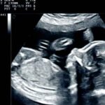
During a prenatal ultrasound, a plastic transducer transmits high-frequency sound waves through your uterus. Sound waves give impulses to a system that creates baby images. Most pregnant women get two ultrasound during pregnancy, but some have more.
What is ultrasound during pregnancy?
Most pregnant women get an ultrasound. It shows your kid in the uterus using sound waves. Ultrasound lets your doctor monitor your baby’s health and development.
Ultrasound is the first time you “see” your baby during pregnancy. If done correctly and your kid is in the right position, you may view his hands, legs, and other body parts. If you don’t want to know if your kid is a boy or a girl, notify your provider!
Most women undergo an ultrasound at 18–20 weeks in their second trimester. Some have a first-trimester ultrasound (early ultrasound) before 14 weeks. Women with asthma and obesity may need more ultrasounds at various times.
Why have an ultrasound during pregnancy?
Your doctor utilizes ultrasonography for:
- Confirm pregnancy.
- Check your baby’s age and growth. This helps your provider calculate your due date.
- Check your baby’s heartbeat, muscle tone, movement, and development.
- Check for pregnancy with twins, triplets, or multiples.
- Ensure your baby is in the head-first position before birth.
- Examine your ovaries and uterus. Your ovaries store eggs.
The doctor utilizes ultrasonography for screening and other tests. Screening determines if your baby is more likely to have a health condition, not if they do. Your doctor may utilize ultrasound:
- Screening for birth problems such as spina bifida and heart disorders. After an ultrasound, your doctor may order diagnostic testing to confirm a birth abnormality. Birth defects are infant health issues. Birth defects alter bodily shape or function. They can affect health, development, and function.
- Prenatal diagnostics such as chorionic villus sampling (CVS) and amniocentesis (amnio) need assistance. CVS tests placenta cells. Your infant gets nutrition from the placenta. Amnio tests amniotic fluid and cells from your baby’s sac.
- Check for pregnancy issues, such as ectopic, molar, and miscarriage.
What routine pregnancy scans are available?
Pregnancy scans are usual. You can choose to have an ultrasound.
Dating scan
Any time between 6 and 14 weeks of pregnancy is suitable for a dating scan. This ultrasound can confirm your pregnancy and approximate the due date. It can clarify how many infants you have and that your baby is growing in your uterus, not ectopic.
Nuchal translucency scan
The nuchal translucency scan, often known as a 12-week scan,’ is normally done around 12 weeks of pregnancy but can be done at 11 weeks to 13 weeks, 6 days. Nuchal translucency is a critical ultrasound measurement. This measurement helps determine your baby’s chromosomal abnormality risk. Additional prenatal screening may include a blood test and ultrasound data to calculate your baby’s risk of chromosomal abnormalities like Down syndrome. Like a dating ultrasound, this scan can assess your baby’s growth, due date, and physical and structural development.
Scan morphology
Between 18 and 22 weeks of pregnancy, a morphology scan (sometimes called a ‘fetal anomaly scan’) is an ultrasound. To estimate gestational age and size, it measures your baby’s organs, including structure and growth. This scan also checks your baby’s heart rate and rhythm and ensures the cervix is long and closed and the placenta is not covering it. Only if asked, this scan may show your baby’s sex, depending on its location.
Is ultrasound during pregnancy risky?
Ultrasound by your doctor is safe for you and your baby. Ultrasound is safer than X-rays since it employs sound waves. For over 30 years, ultrasonography providers have discovered no dangers.
If your pregnancy is healthy, ultrasonography can rule out most issues, but not all. It may miss birth problems. Routine ultrasounds may misdiagnose birth defects. While follow-up testing usually confirms the infant is healthy, false alarms can worry parents.
Conclusion
Ultrasounds are generally accurate at estimating a baby’s growth, but they sometimes underestimate or overestimate weight, leading to unneeded C-sections or premature deliveries. If your doctor suggests more than two ultrasounds while you’re pregnant, request them to confirm their medical necessity.